
January 31
In the second half of The Double Helix and in The Code Breaker, there begin to be some references to the structures of DNA and protein, to the genetic code, to mutations and their effects and, eventually, to mechanisms of CRISPR action. Full understanding of these technical discussions is not necessary to grasp and discuss the main issues raised by the books. However, in case you may be interested in further understanding some of these issues, there are links below to some resources that may help.
Here are some links to resources that explain basic aspects of DNA biology and structure.
YourGenome.org is a well-organized, nicely illustrated introduction to “your genome”. Relatively basic and very accessible, this is an excellent starting resource.
https://www.yourgenome.org/facts/what-is-dna
DNA From The Beginning is “An animated primer of 75 experiments that that made modern genetics” produced by the Cold Spring Harbor Laboratory DNA Center. This website is a couple decades old and consequently seems a little rusty, but it is very well organized and illustrates important milestones in genetics and molecular biology with animations, videos and brief descriptive material.
The Khan Academy has excellent segments on DNA discovery, structure and function:
https://www.khanacademy.org/science/biology/dna-as-the-genetic-material#dna-discovery-and-structure
The Learn.Genetics website was created by the outstanding genetics unit at the University of Utah. This site is a little more basic that the two previous, but is illustrated with outstanding short and easy-to-understand videos.
https://learn.genetics.utah.edu/content/basics/
https://learn.genetics.utah.edu/content/basics/dna
The HHMI Biointeractive Video titled The DNA Double Helix Discovery is a very professional and engaging account of the discovery of DNA’s structure. (17 minutes)
https://www.youtube.com/watch?v=1vm3od_UmFg
The video linked below is a lecture given by Eric Lander (whom we will memorably encounter in The Code Breaker), describing the key experiments demonstrating that DNA, not protein, is the genetic material (Avery, MacLeod and McCarty, 1944). Although 45 minutes long, the lecture is lucid and gripping! Take a look!
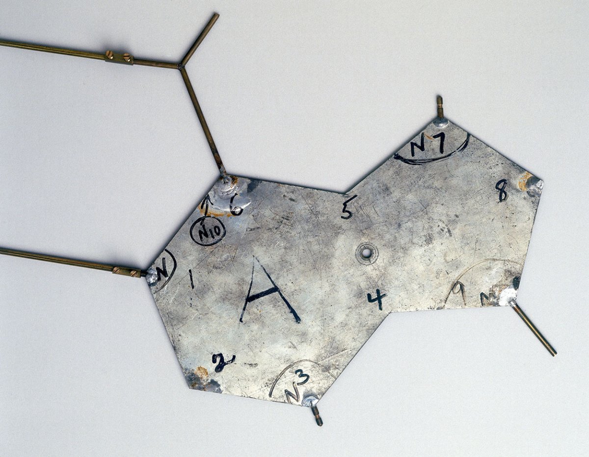
Template from Francis Crick and James Watson’s molecular model of DNA from 1953. Image credit: Wikimedia Commons.
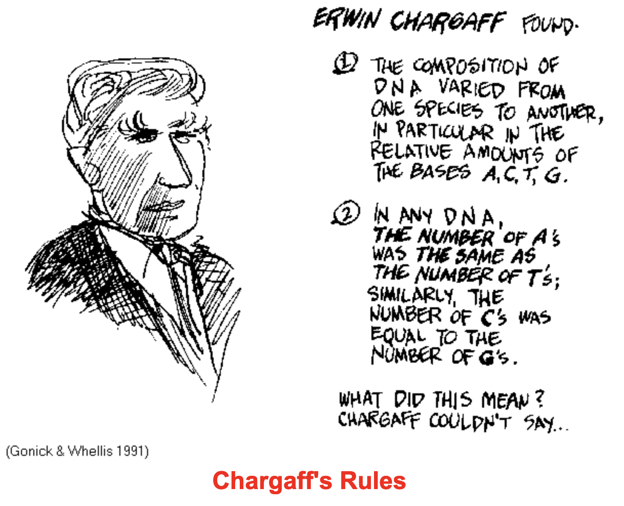
Edwin Chargaff's careful investigation of the base composition of DNA from different organisms provided a strong hint that helped solve the structure of DNA.


The left panel shows the structures of the four bases ans of a nucleotide. The right panel shows the phosphodiester bonds linking nucleotides to form one-half of a double helix.
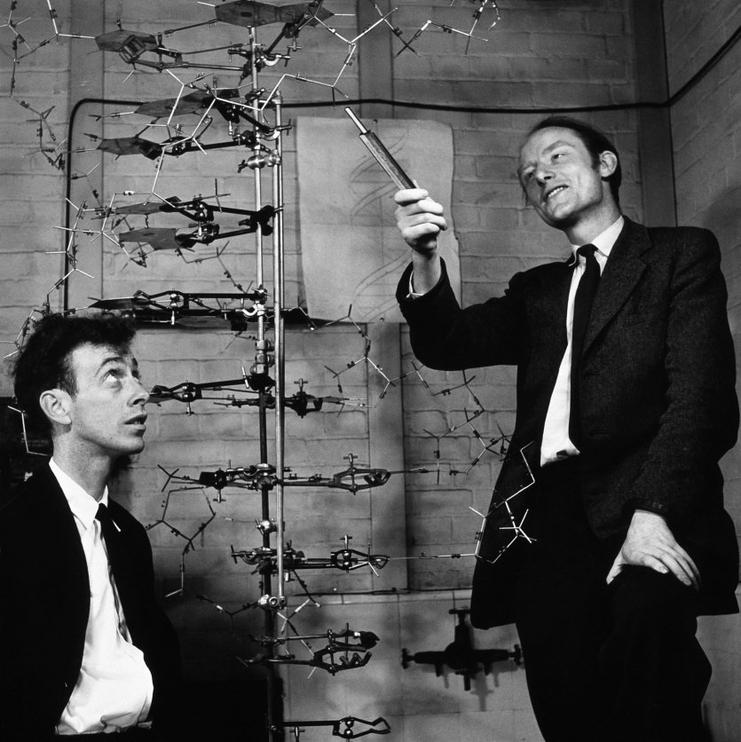
James Watson and Francis Crick in 1953. Credit: A. Barrington Brown/Science Photo Library

This panel shows a short segment of a double helix. Adenine and thymine (A and T) are held together by two hydrogen bonds (dotted lines), while guanine and cytosine (G and C) are held together by three hydrogen bonds. The two chains are oriented in an anti-parallel configuration

The Eagle - the pub near the old Cavendish Laboratory, where Watson and Crick frequently ate lunch....and where Crick, on February 28, 1953 announced to the patrons that he and Watson had just discovered the secret of life!

Outside the Eagle, this plaque commemorates Watson and Crick's accomplishment - it was unveiled by James Watson at a ceremony on the 50th anniversary of their discovery.

(L-R) Maurice Wilkins (Physiology or Medicine), M. Perutz (Chemistry), Francis Crick (Physiology or Medicine), J. Steinbeck (Literature), James Watson (Physiology or Medicine), J. Kendrew (Chemistry).
Related readings
Here’s a very objective and readable review of Rosalind Franklin’s contributions and interactions with the other DNA researchers.
Here is the famous 1953 Nature paper describing the proposed double helix structure for DNA.
See this article for a good, simple description of the use of x-ray diffraction for determining the structure of molecules that have been crystallized. As illustrated below, patterns obtained for proteins are complex and difficult to interpret!

The left panel shows the famous “Photo 51” x-ray diffraction picture of B-form DNA. For comparison, the x-ray diffraction pattern on the right is derived from a small protein (egg white lysozyme).
Why is the pattern for DNA so much simpler than that for even a small protein? Lysozyme is a small protein, but still gives a complicated pattern because it is composed of 129 amino acids, each of which would have between 10 and 25 atoms…thus there would be something like 2,500 atoms in lysozyme - each of which would be regularly repeated in a crystal of lysozyme. Although a double-helix DNA strand would contain many more atoms than lysozyme, I believe it gives a simpler pattern because the only features of DNA that are repeated in a regular way in crystals would be the components of the two sugar-phosphate backbones; the individual bases are not repeated in a regular manner and so would not generate consistent reflections.